Have you been inactive for a while and are now ready to get back into shape? Returning to fitness after a period of inactivity can be daunting, but with the right approach, it is achievable.
Life can sometimes get in the way, causing us to neglect our fitness routines. Being inactive for an extended period can make it difficult to regain our previous fitness level, whether it’s due to injury, illness, or other obligations.
Returning to shape after inactivity requires patience, persistence, and a well-designed plan. It’s important to start slowly and gradually increase your workouts’ intensity and duration. By avoiding common mistakes and following the right strategies, you can return to fitness and regain strength and stamina.
Movement affected human evolution. Foraging for food, long-distance travel, and battling off predators were all necessary for survival. Many biological systems have evolved into this way of living and they must be active to work optimally.
Regular physical activity encourages the creation of natural substances such as hormones, enzymes, and antioxidants. Products that regulate the processes of development and repair. Sedentary lifestyles disrupt these essential functions.
Inactivity promotes the creation of several compounds, such as excessive inflammation and oxidative stress as well as possible cancer cell development. High amounts of these compounds may be harmful.
Physical inactivity is a key risk factor for health.[1] Being sedentary raises the risk of cancer, abnormal blood work, diabetes, cardiovascular disease, obesity, chronic fatigue and respiratory disease.[2]
The time it takes to go from sedentary to fit will vary depending on individual factors such as age, occupation, previous fitness levels, and health conditions.
Regular exercise will provide you with more significant fitness and lifestyle benefits over time. Individuals can vary however, you can see differences after 6 to 8 weeks.[3] You may completely transform your health and fitness in 3 to 4 months, which is approximately the same time frame as strength-specific exercises.
Generally speaking, it can take several weeks to months to become fit after a prolonged period of inactivity, working with a trained professional can help to ensure you are doing what’s best for you and your body without injuring yourself.
Absolutely. It’s never too late to start making positive changes in your lifestyle and reversing the damage caused by a sedentary lifestyle. Even if you’ve been inactive for years, it’s still possible to improve your physical and mental health with regular exercise.
All it takes is a little dedication and planning. Start by setting realistic goals for yourself – such as exercising 30 minutes a day, 3 times a week – and work your way up. Soon enough, you’ll be reaping the rewards of improved health!
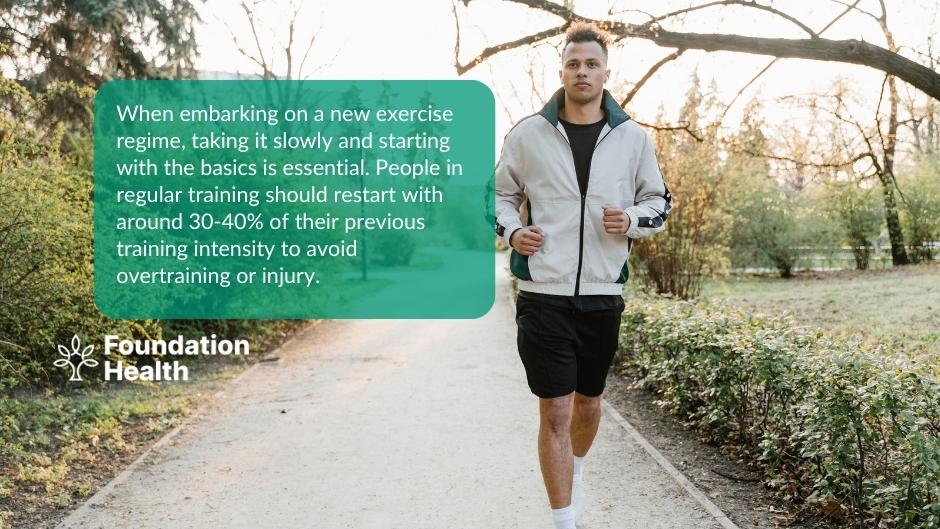
When embarking on a new exercise regime, taking it slowly and starting with the basics is essential. People in regular training should restart with around 30-40% of their previous training intensity to avoid overtraining or injury.
This is more beneficial than beginning again at full force. Suddenly increasing the level of activity could lead to an overload of stress on the body resulting in adverse side effects like fatigue, muscle tightness and strain or injury.
To reduce the chance of this occurring, it is essential to include a warm-up and cool-down period into your routine before and after exercise. An effective warm-up should last around 10 minutes, typically involving stretches and dynamic movements that raise your heart rate slightly.[6]
Then, during aerobic training, focus on low-intensity endurance exercises such as jogging or cycling for the first four weeks before attempting any high-intensity training program.
Strength training should also be started gradually by increasing reps rather than weight for 2-3 weeks until you build enough strength to return to higher weights.
Regardless of the type of training performed, aim to regain your previous level after 1 month, allowing gradual progress without putting too much pressure on yourself from day one.
Gentle exercise classes such as pilates and yoga can also be a fantastic way to ease back into exercise. These modalities focus on correct positioning and alignment to ensure the correct muscle groups are working effectively. Group classes or exercising with a loved one can be a great way to hold you accountable as well as make exercising more enjoyable.
Regular exercise strengthens the cardiac muscles (the heart) reducing the risks of heart attacks and increases blood circulation, promoting oxygen levels in the body.[7] Exercise also increases energy levels, improves sleep, lowers cortisol, and acts as a natural mood booster. Exercise has been proven to assist with conditions such as depression and anxiety as well as helping reduce the risk of type 2 diabetes, stroke, high blood pressure, and reduces the likelihood of falls in the elderly population.
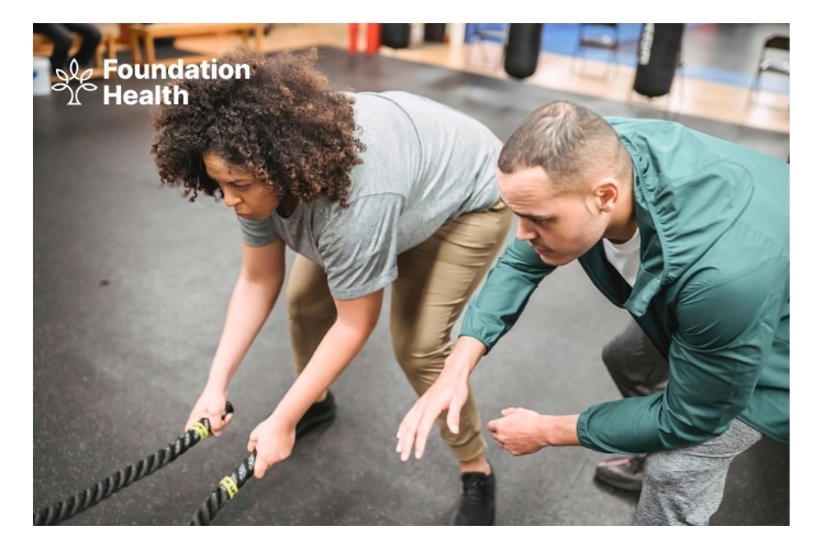
Returning to fitness after a period of inactivity can be daunting, but it is achievable with the right approach and dedication. Start slowly and gradually increase your workouts’ intensity and duration.
Additionally, set realistic goals, take breaks to recover from workouts and find an activity you enjoy to keep you motivated.
Ensuring you look after your body during this transition period is also vital. Taking time to rest and recover from workouts is essential for preventing injury or burnout.
Furthermore, regular osteopathic treatments can help manage any existing or potential injuries. Osteopathy assists with improving joint mobility, reducing pain and ensuring correct posture throughout the rehabilitation process.
Book an appointment at Foundation Health for osteopathic treatment that will assist in managing your return to fitness.
1. Richard Patterson; Eoin McNamara; Marko Tainio; Thiago Hérick de Sá; Andrea D. Smith; Stephen J. Sharp; Phil Edwards; James Woodcock; Søren Brage; Katrien Wijndaele (2018). Sedentary behaviour and risk of all-cause, cardiovascular and cancer mortality, and incident type 2 diabetes: a systematic review and dose response meta-analysis. European Journal of Epidemiology. https://doi.org/10.1007/s10654-018-0380-1 ↩
2. Aviroop Biswas; Paul I. Oh; Guy E. Faulkner; Ravi R. Bajaj; Michael A. Silver; Marc S. Mitchell; David A. Alter (2015). Sedentary Time and Its Association With Risk for Disease Incidence, Mortality, and Hospitalization in Adults. Annals of Internal Medicine. https://doi.org/10.7326/m14-1651 ↩
3. Felipe Damas; Stuart M. Phillips; Cleiton A. Libardi; Felipe C. Vechin; Manoel E. Lixandrão; Paulo R. Jannig; Luiz A. R. Costa; Aline V. Bacurau; Tim Snijders; Gianni Parise; Valmor Tricoli; Hamilton Roschel; Carlos Ugrinowitsch (2016). Resistance training‐induced changes in integrated myofibrillar protein synthesis are related to hypertrophy only after attenuation of muscle damage. The Journal of Physiology. https://doi.org/10.1113/jp272472 ↩
4. Ron Borde; Tibor Hortobágyi; Urs Granacher (2015). Dose–Response Relationships of Resistance Training in Healthy Old Adults: A Systematic Review and Meta-Analysis. Sports Medicine. https://doi.org/10.1007/s40279-015-0385-9 ↩
5. Leslie H. Willis; Cris A. Slentz; Lori A. Bateman; A. Tamlyn Shields; Lucy W. Piner; Connie W. Bales; Joseph A. Houmard; William E. Kraus (2012). Effects of aerobic and/or resistance training on body mass and fat mass in overweight or obese adults. Journal of Applied Physiology. https://doi.org/10.1152/japplphysiol.01370.2011 ↩
6. Courtney J. McGowan; David B. Pyne; Kevin G. Thompson; Ben Rattray (2015). Warm-Up Strategies for Sport and Exercise: Mechanisms and Applications. Sports Medicine. https://doi.org/10.1007/s40279-015-0376-x ↩
7. Matthew A. Nystoriak; Aruni Bhatnagar (2018). Cardiovascular Effects and Benefits of Exercise. Frontiers in Cardiovascular Medicine. https://doi.org/10.3389/fcvm.2018.00135 ↩
Have you ever tweaked your ankle while running or felt a sharp pain in your knee? If so, you’re not alone: running can be hard on the body, and injuries are common.[1] But don’t worry – help is available.
Osteopathy is a manual therapy that can help with running injuries. They’ll help get you back on your feet – and running – in no time.
Running injuries can occur for various reasons, including:
Most runners go over on their ankles, sprain them, and are back to square one soon after. Ankle pain usually occurs when you twist or fall on your ankle.[3]
The majority of sprains will heal with rest, ice, and elevation.[3] However, it’s still a good idea to see an osteopath for a sports massage, recovery procedures, and strengthening exercises to help the ankle heal faster.[3]
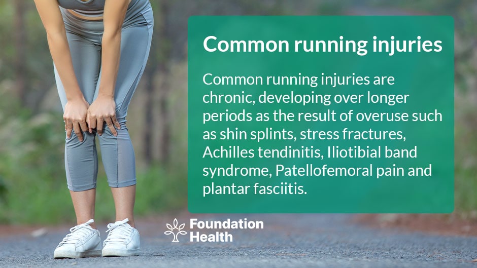
This injury, also known as shin splints, is caused by excessive tibial strain.[2] Runners with this condition typically experience pain anywhere along their inner shin.
Pain near the kneecap, also known as “runner’s knee”, is commonly associated with long-distance running.[4]
When you run, this muscle is constantly under strain and stress; it usually feels tight and stiff, but it can become increasingly worse.
A condition in which the plantar fascia ligament, which is located between the toes and the heel, becomes inflamed after prolonged training on hard terrain.[2]
Pulling your hamstring can be excruciatingly painful and can occur at any point along the muscle. Pulled hamstrings can occur due to various factors, including fatigue, running at a strained pace, and issues with other muscles that place additional strain on your hamstring.
This injury, also known as “jumper’s knee,” involves damage to the patella tendon, which is located just below the kneecap. It is commonly associated with long periods of running.
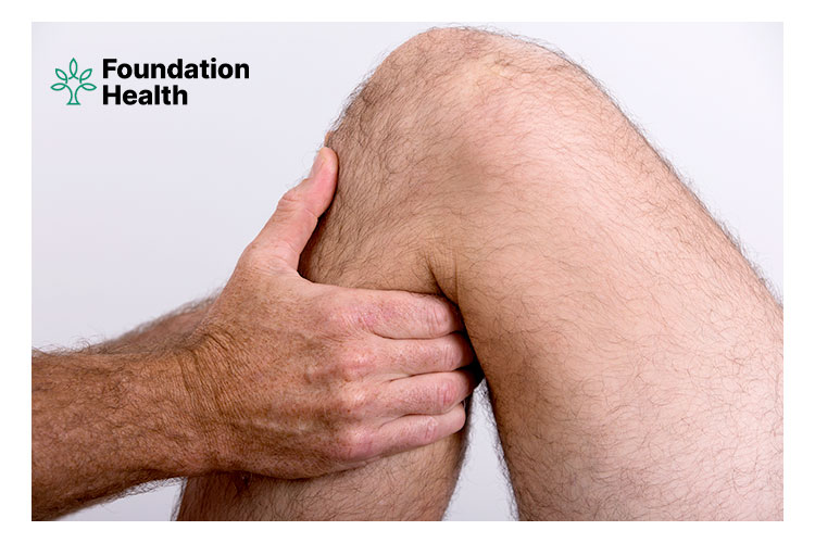
Increasing joint mobility and range of motion through gentle manipulation and articulation.[3]
This procedure focuses on increasing blood flow and releasing and relaxing muscles through deep pressure massage and trigger point therapy.
This essentially entails assessing your gait, both while walking and while running. This evaluation of your movement will include an examination of your back, hips, knees, and feet and how they are affected while running.
Your osteopath will recommend specific stretches and exercises for injury recovery and day-to-day training methods.[4]
Runners can benefit significantly from osteopathy. An osteopath will evaluate your running technique, shoe wear patterns, and training load in conjunction with an osteopathic musculoskeletal examination to help form a picture of why the runner has become injured. The injury will then be treated by an osteopath using a combination of soft tissue treatment, joint articulation or manipulation, dry needling (acupuncture), and ergonomic, rehabilitation, and training advice.
Osteopaths are trained to use their knowledge of biomechanics to identify areas of your body that are not functioning optimally and work on them to improve your health and performance.
If you are new to running, you may be more likely to require treatment for runner’s injuries.[1] If you increase the length or intensity of your running routine, you may be at greater risk of running injuries.[1]
Before you end up needing treatment for running injuries, it might be worth paying a visit to your osteopath. An osteopathic evaluation is your best bet for avoiding running injuries.
This is because we recognise that running is an excellent way to get in shape. It improves cardio-vascular fitness and muscle tone and can even improve your mood![5] As a result, we strongly encourage you to participate. However, running can be dangerous, even for marathon runners.
Whether you run marathons, jog for general fitness, or use running as a training aid for other sports, there is always room for improvement in performance, injury prevention, and rehabilitation.
Osteopathy provides numerous long-term benefits to the body, mind, and spirit. Osteopathy can be used as a preventative measure, allowing athletes and runners to avoid similar injuries in the future.
Osteopathy’s primary goal is to get your entire body moving correctly. This makes your body more efficient, allowing you to waste less energy and redirect it towards healing and recovery. In the end, this means fewer injuries, less time off, and improved performance.
If you’re struggling with a running injury, book an appointment with one of our osteopaths at Foundation Health to diagnose and rehabilitate your injury.
1. Solvej Videbæk; Andreas Moeballe Bueno; Rasmus Oestergaard Nielsen; Sten Rasmussen (2015). Incidence of Running-Related Injuries Per 1000 h of running in Different Types of Runners: A Systematic Review and Meta-Analysis. Sports Medicine. https://doi.org/10.1007/s40279-015-0333-8 ↩
2. Bruno R. da Costa; Edgar Ramos Vieira (2009). Risk factors for work‐related musculoskeletal disorders: a systematic review of recent longitudinal studies. American Journal of Industrial Medicine. https://doi.org/10.1002/ajim.20750 ↩
3. Gwendolyn Vuurberg; Alexander Hoorntje; Lauren M Wink; Brent F W van der Doelen; Michel P van den Bekerom; Rienk Dekker; C Niek van Dijk; Rover Krips; Masja C M Loogman; Milan L Ridderikhof; Frank F Smithuis; Sjoerd A S Stufkens; Evert A L M Verhagen; Rob A de Bie; Gino M M J Kerkhoffs (2018). Diagnosis, treatment and prevention of ankle sprains: update of an evidence-based clinical guideline. British Journal of Sports Medicine. https://doi.org/10.1136/bjsports-2017-098106 ↩
4. Kay M Crossley; Marienke van Middelkoop; Michael J Callaghan; Natalie J Collins; Michael Skovdal Rathleff; Christian J Barton (2016). 2016 Patellofemoral pain consensus statement from the 4th International Patellofemoral Pain Research Retreat, Manchester. Part 2: recommended physical interventions (exercise, taping, bracing, foot orthoses and combined interventions). British Journal of Sports Medicine. https://doi.org/10.1136/bjsports-2016-096268 ↩
5. Eduard Alentorn-Geli; Kristian Samuelsson; Volker Musahl; Cynthia L. Green; Mohit Bhandari; Jón Karlsson (2017). The Association of Recreational and Competitive Running With Hip and Knee Osteoarthritis: A Systematic Review and Meta-analysis. Journal of Orthopaedic & Sports Physical Therapy. https://doi.org/10.2519/jospt.2017.7137 ↩
The most common causes of muscular pain and aching muscles are overuse, increased tension, and trauma. All of which can occur due to overtraining.
Muscle strains, also known as pulled muscles, occur when the muscle is overstretched, causing fibres to tear.[3] This can happen if you are tired, have poor flexibility, perform a movement that your body is not used to or perform repetitive movements/activities.
If pain relievers, anti-inflammatory medications, and rest haven’t provided you with long-term relief from muscular aches and pains, it’s time to see an osteopath.
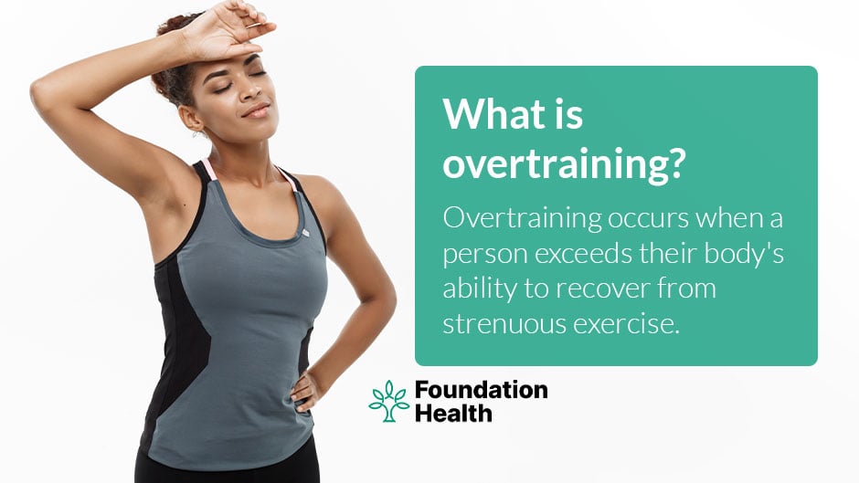
Overtraining occurs when a person is unable to recover from strenuous activity.[1] Performance declines and plateaus as a result of overtraining.[1] This is the inability to perform consistently at a certain level or train at a load that exceeds a person’s capacity for recovery. People who overtrain stop progressing and can even lose strength and fitness.[1] Athletes who overtrain also experience chronic fatigue, burnout, and overstress.[1]
The majority of people will experience pain, weakness, and limited mobility.[3] Swelling and bruising may occur depending on the severity of the injury.[3] Surgery may be required if the injury is severe.[3] Most muscle strains are mild to moderate in severity and heal within 2-4 weeks; however, more painful tears can take months to heal.[3]
Effective training and injury prevention require active warm-ups and movement preparation.[4] Avoiding a warm-up leaves you vulnerable to injury. Activate the area you are working on for at least 10 minutes to stimulate the blood flow. This will get your body moving!
It’s advisable to begin stretching with what’s known as “dynamic stretching.” This involves stretches that incorporate movement. Dynamic stretching prepares your body for what’s to come by gently increasing your range of motion and raising your heart rate while also getting your blood flowing.[5]
Post-exercise is the best time to perform “static stretching” in areas that may feel tight—these are commonly known stretches that you hold in one position for 15-30 seconds. Since your muscles are already warm after your workout, stretching will benefit them more than if you did it when they are cold.
Sleep is most likely the most underutilised recovery tool.[6] You are not recovering if you are not sleeping.[6] Relaxation time is significant for optimal recovery, so make it a priority to rest when you can.
Training will vary depending on the exercises you choose and how you build it up. Those who have been sedentary for a long time will need to start much slower than those who wish to advance their previous training programs.[7]
Excessive training should be avoided. You must manage the time between sessions so your body can get adequate rest. Whether you are trying to gain muscle or lose weight, overtraining will put your body under stress, leading to a breakdown of your body’s repair system.
As a general rule, you should increase your load by 10% a week and allow for adequate rest time between hard training sessions. Most experts agree that this should be at least 24 hours, if not up to 48 hours, and never underestimate the value of a rest day![1]
An osteopath can help with muscle pain.[8] Osteopathy can help by manipulating a patient’s muscles, tendons, and ligaments.[8] This causes tight muscles to relax, circulation to improve, and trapped ligaments and tendons to be released. During soft tissue release, by assisting in the alignment of your joints and smoothing out areas of scar tissue and other muscular irregularities, the muscular pain cycle is broken, the healing process is accelerated, and muscular balance is restored.
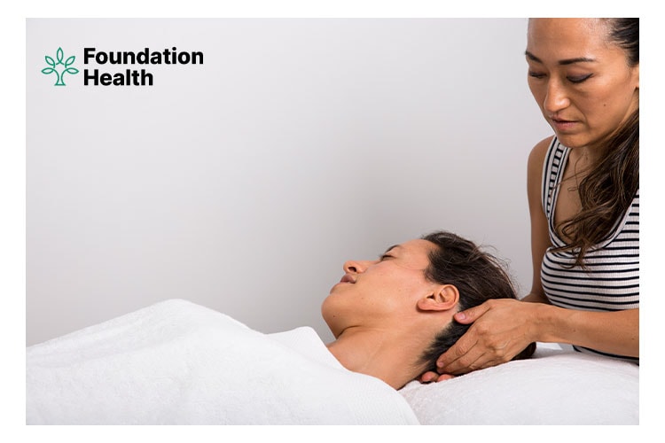
Overtraining can be assessed, diagnosed, and managed using osteopathy. Gym-related injuries and overtraining account for the vast majority of the patients we see as osteopaths daily.
Overtraining can impair performance and lead to long-term psychological and physical issues.
If you experience sharp, intense pain while working out in the gym, this is your body’s way of alerting you that something is wrong.[9] It either means you’ve already been injured or that one is on the way.
Please book an appointment with one of our osteopaths at Foundation Health to diagnose your injury and plan ways to train around it!
1. Romain Meeusen; Martine Duclos; Carl Foster; Andrew Fry; Michael Gleeson; David Nieman; John Raglin; Gerard Rietjens; Jürgen Steinacker; Axel Urhausen (2013). Prevention, Diagnosis, and Treatment of the Overtraining Syndrome. Medicine & Science in Sports & Exercise. https://doi.org/10.1249/mss.0b013e318279a10a ↩
2. Tim J Gabbett (2016). The training—injury prevention paradox: should athletes be training smarter and harder?. British Journal of Sports Medicine. https://doi.org/10.1136/bjsports-2015-095788 ↩
3. Lauren N. Erickson; Marc A. Sherry (2017). Rehabilitation and return to sport after hamstring strain injury. Journal of Sport and Health Science. https://doi.org/10.1016/j.jshs.2017.04.001 ↩
4. Carolyn A Emery; Thierry-Olivier Roy; Jackie L Whittaker; Alberto Nettel-Aguirre; Willem van Mechelen (2015). Neuromuscular training injury prevention strategies in youth sport: a systematic review and meta-analysis. British Journal of Sports Medicine. https://doi.org/10.1136/bjsports-2015-094639 ↩
5. David G. Behm; Anthony J. Blazevich; Anthony D. Kay; Malachy McHugh (2016). Acute effects of muscle stretching on physical performance, range of motion, and injury incidence in healthy active individuals: a systematic review. Applied Physiology, Nutrition, and Metabolism. https://doi.org/10.1139/apnm-2015-0235 ↩
6. Neil P Walsh; Shona L Halson; Charli Sargent; Gregory D Roach; Mathieu Nédélec; Luke Gupta; Jonathan Leeder; Hugh H Fullagar; Aaron J Coutts; Ben J Edwards; Samuel A Pullinger; Colin M Robertson; Jatin G Burniston; Michele Lastella; Yann Le Meur; Christophe Hausswirth; Amy M Bender; Michael A Grandner; Charles H Samuels (2020). Sleep and the athlete: narrative review and 2021 expert consensus recommendations. British Journal of Sports Medicine. https://doi.org/10.1136/bjsports-2020-102025 ↩
7. Fiona C Bull; Salih S Al-Ansari; Stuart Biddle; Katja Borodulin; Matthew P Buman; Greet Cardon; Catherine Carty; Jean-Philippe Chaput; Sebastien Chastin; Roger Chou; Paddy C Dempsey; Loretta DiPietro; Ulf Ekelund; Joseph Firth; Christine M Friedenreich; Leandro Garcia; Muthoni Gichu; Russell Jago; Peter T Katzmarzyk; Estelle Lambert; Michael Leitzmann; Karen Milton; Francisco B Ortega; Chathuranga Ranasinghe; Emmanuel Stamatakis; Anne Tiedemann; Richard P Troiano; Hidde P van der Ploeg; Vicky Wari; Juana F Willumsen (2020). World Health Organization 2020 guidelines on physical activity and sedentary behaviour. British Journal of Sports Medicine. https://doi.org/10.1136/bjsports-2020-102955 ↩
8. Donatella Bagagiolo; Debora Rosa; Francesca Borrelli (2022). Efficacy and safety of osteopathic manipulative treatment: an overview of systematic reviews. BMJ Open. https://doi.org/10.1136/bmjopen-2021-053468 ↩
9. Adrienne E. Dubin; Ardem Patapoutian (2010). Nociceptors: the sensors of the pain pathway. Journal of Clinical Investigation. https://doi.org/10.1172/jci42843 ↩
Brazilian Jiu-Jitsu (BJJ) is a grappling-based martial art that can cause injuries during training and competition.
As submission wrestling, grappling comprises chokes/strangles and joint locks. Furthermore, BJJ contains aggressive takedowns from standing and self-defense techniques to force your opponent to submit or “flee” the situation in self-defense.
Research has determined that 78% of injuries sustained in BJJ contests are orthopaedic injuries, many of which necessitate medical treatment or surgical intervention.[1]
Overuse and direct contact, a lack of conditioning, and soft tissue insufficiency are significant causes of Brazilian Jiu-Jitsu practitioners’ injury.[2]
The dynamics of the BJJ fight, particularly the techniques, create overloads in the joints (mainly in the joints of the upper and lower limbs) by pressing them to the extreme range of motion, frequently exceeding the tolerance of the tissues, which can result in microtrauma and, as a result, discomfort and pain.[3]
This overload might happen during a single incident of executing a specific movement or due to repetitive drills. Knee and shoulder joints, ligament injuries and dislocations are the most common, with takedowns, foot locks and arm locks being the techniques as the cause of these injuries.[2]
For example, there are two typical BJJ injuries caused by the “kimura” jiu-jitsu technique. A “kimura” is performed by securing an opponent’s wrist, then internally rotating their shoulder and elbow by applying internal rotation pressure to the secured wrist and arm.
The risk of injury from using a “kimura” arises when both the elbow and shoulder are manipulated, potentially harming several anatomic tissues.[1] This typically leads to a grade I AC separation in the shoulder and an MCL sprain in the elbow.
Traumatic finger polyarthrosis, which is essentially a wear and tear injury to the joints and ligaments in the fingers, is another common injury in BJJ.[4] It frequently occurs as a result of ignoring minor injuries over time. It can result in finger joint deformity and pain, and weakness in the hands.[4]
It is beneficial to perform neuromuscular and proprioception training for at least 30 minutes to prevent injuries due to their ability to prepare and develop the body for the specific demands of the sport. Furthermore, neuromuscular and proprioception training can be done as a drill, which would not be detrimental because it is a typical activity in BJJ training.
The most practical method to avoid neck injuries in BJJ is to avoid fully stacked positions with your weight on your neck; instead, attempt to keep your weight on your shoulders.
Ensure to strengthen the muscles surrounding your knee and avoid exerting lateral tension on your knee joint for optimal knee health.
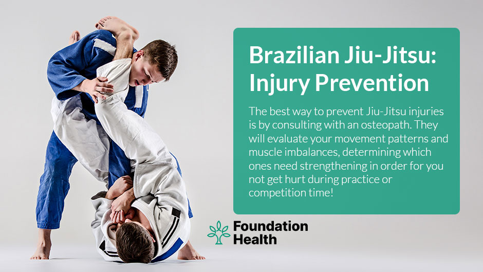
The best course of action is to consult with an osteopath to evaluate your movement patterns and muscle imbalances to determine how to strengthen susceptible muscles and increase joint mobility to circumvent common Brazilian Jiu-Jitsu injuries.
Not every grappler will be affected by cauliflower ear; it is highly variable. Cauliflower ear occurs when an ear injury causes blood to accumulate and pool beneath the skin, also known as a perichondrial hematoma.[5] The pooling blood causes the skin to feel spongy, rubbery, and lumpy.
A perichondrial hematoma is typically not any cause for concern. This can, however, permanently change the appearance of the ear. As the cartilage dies, a person’s ear can flop over.
A medical professional should remove the fluid as soon as possible to reduce the likelihood of clot formation.[5] In most cases, the fluid is drained using a wide-bore needle in a procedure known as aspiration.[5] This fluid may contain blood clots, preventing further obstructions.
Constant compression is required to keep the injury from filling up with fluid again.[5] Compression also prevents the skin from ripping out more blood vessels by keeping it close to the cartilage. You should avoid further BJJ training and any other situations where ear trauma is likely to occur again.[5]
In BJJ, hand and finger injuries are practically unavoidable. Taping your fingers provides extra support to the joints and tendons of your fingers, allowing you to continue training with less pain. As long as you continue to cause stress on your fingers that caused your injury, your fingers won’t heal.
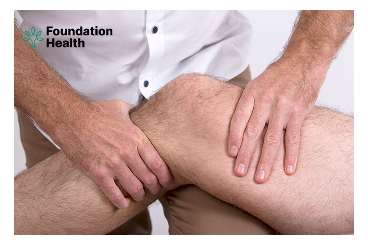
A catastrophic injury is one of the simplest ways to end your Brazilian Jiu-Jitsu career. It is possible to train after a BJJ injury, but avoiding these injuries should be everyone’s goal! Knowing the most common sites of injury and how to avoid them is critical for anyone hoping to train Jiu-Jitsu for a long time.
If you are suffering from pain due to Brazilian Jiu-Jitsu, book an appointment with one of our expert osteopaths at Foundation Health for a professional assessment today!
1. James F. Scoggin; Georgiy Brusovanik; Byron H. Izuka; Eddy Zandee van Rilland; Olga Geling; Seren Tokumura (2014). Assessment of Injuries During Brazilian Jiu-Jitsu Competition. Orthopaedic Journal of Sports Medicine. https://doi.org/10.1177/2325967114522184 ↩
2. Maximilian Hinz; Benjamin D. Kleim; Daniel P. Berthold; Stephanie Geyer; Christophe Lambert; Andreas B. Imhoff; Julian Mehl (2021). Injury Patterns, Risk Factors, and Return to Sport in Brazilian Jiu Jitsu: A Cross-sectional Survey of 1140 Athletes. Orthopaedic Journal of Sports Medicine. https://doi.org/10.1177/23259671211062568 ↩
3. Brad A. Petrisor; Gina Del Fabbro; Kim Madden; Moin Khan; Jeff Joslin; Mohit Bhandari (2019). Injury in Brazilian Jiu-Jitsu Training. Sports Health: A Multidisciplinary Approach. https://doi.org/10.1177/1941738119849112 ↩
4. P. Strasser; M. Hauser; H. J. Häuselmann; B. A. Michel; A. Frei; G. Stucki (1997). Traumatische Fingerpolyarthrose bei Judo-Sportlern: Eine Verlaufsuntersuchung (Development of finger-joint osteoarthritis in Judo). Zeitschrift für Rheumatologie. https://doi.org/10.1007/s003930050048 ↩
5. Brit Long; Jessica Mason; Rachel E. Bridwell; Michael Gottlieb (2025). Managing Auricular Hematoma: An Emergency Medicine Narrative Review. The Journal of Emergency Medicine. https://doi.org/10.1016/j.jemermed.2024.08.021 ↩
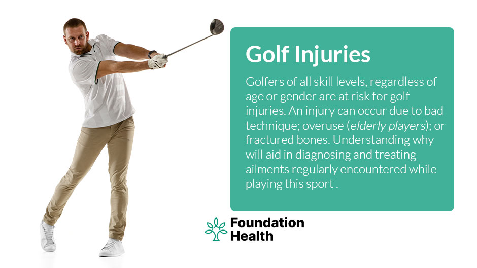
Many people perceive golf as a low-intensity physical sport that does not provide a significant risk of injury. However, the explosive nature of the swing can put a tremendous amount of stress on the body.
Golf injuries are prevalent among golfers of all skill levels, regardless of age or gender.[1] An injury can occur due to bad technique, overuse, or a direct blow to the body.[2] Golfing can cause various types of injury, including bone fractures, ankle sprain, elbow strain, spinal issues, knee pain, hip pain, back pain, and wrist sprains.
Understanding why will aid in the timely diagnosis and treatment of ailments regularly encountered while playing golf.
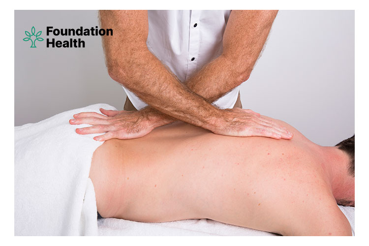
When wrist discomfort and swelling initially emerge, the best course of action is to rest and apply ice to reduce swelling. Heat applied to the affected area might help reduce pain and stiffness. A compression wrap, wrist brace or splint may also be beneficial. If pain persists, then consult an osteopath for an assessment and treatment.
Golf injuries can be prevented by improving swing mechanics, participating in golf-specific training and conditioning programs, purchasing adequately fitted equipment, avoiding long practice sessions, always performing a warm-up routine before practice and play, and golf-specific stretching regularly.
Preventing golf injuries is better than having to recover from them. The best way to avoid these common golf injuries is to be physically prepared to play.
To be effective, rehabilitation and prevention programs need to address both movement patterns and muscle imbalances as well as technical swings.
If you are suffering from pain due to playing golf, book an appointment with one of our expert osteopaths at Foundation Health for a professional assessment today!
1. Thomas R Williamson; Robert S Kay; Patrick G Robinson; Andrew D Murray; Nicholas D Clement (2024). Epidemiology of musculoskeletal injury in professional and amateur golfers: a systematic review and meta-analysis. British Journal of Sports Medicine. https://doi.org/10.1136/bjsports-2023-107324 ↩
2. Andrew McHardy; Henry Pollard; Kehui Luo (2006). Golf Injuries. Sports Medicine. https://doi.org/10.2165/00007256-200636020-00006 ↩
3. Roger Hawkes; Phil O’Connor; Doug Campbell (2013). The prevalence, variety and impact of wrist problems in elite professional golfers on the European Tour. British Journal of Sports Medicine. https://doi.org/10.1136/bjsports-2012-091917 ↩
4. Patrick G Robinson; Iain R Murray; Andrew D Duckworth; Roger Hawkes; Danny Glover; Nigel R Tilley; Rob Hillman; Christopher W Oliver; Andrew D Murray (2018). Systematic review of musculoskeletal injuries in professional golfers. British Journal of Sports Medicine. https://doi.org/10.1136/bjsports-2018-099572 ↩
5. Hwang-Woon Moon; Jun-Su Kim (2023). Golf-related sports injuries of the musculoskeletal system. Journal of Exercise Rehabilitation. https://doi.org/10.12965/jer.2346128.064 ↩

Hello readers! We hope you’ve had an enjoyable Christmas and sent 2020 off with the bang it deserved. We’re kicking things off with a blog about neck pain relating to small joints in our neck known as facet joints. Are you waking up to 2021 with a pain in your neck? You might have had one too many sleeps in the armchair over the festive period. And maybe the exercise dropped off a bit as focus changed to family get-togethers and binging in front of the TV after an exhausting year. Never fear, we’ve got your back (oops… we mean neck!)
Facet joints are small joints in the neck, formed between bony parts of two adjacent vertebrae[1]. With a few exceptions, you can find a pair of facet joints at each level of the spine: one on the left, one on the right.[1] These joints, along with the disc connection between vertebrae, are responsible for allowing and restricting movements of the spine, depending on what region of the spine you are looking at.[1] For example, the facet joints in the neck are orientated to allow a relatively wide range of motion in all planes of movement… Flexion and extension, rotation, and side-bending (lateral flexion).[1] When we look over our shoulder to check our blind spot in the car, we are mainly using movement in our neck to get there.
If you move to the low back region of the spinal column, the facet joints are orientated in a slightly different way, allowing plenty of flexion and extension, but minimal rotation.[1] This allows us to bend our bodies forwards and backwards easily.
This condition is pretty self-explanatory from its name. A facet lock is a facet joint that is ‘locked’ or severely restricted in movement. This type of joint is what we call a synovial joint. This means it’s a joint that is held together by a joint capsule and is filled with a lubricating fluid, known as synovial fluid.[1] A facet joint tends to lock when it has been overloaded with excessive forces acting upon it. This tends to occur over time and results in a ‘straw that breaks the camel’s back’ moment. It can also happen following a quick jerking movement of the neck, where a sudden large force is placed upon the joints and it is too much for them to bear.[1] The tissue around the joints, including the overlying muscles which drive the movement stiffen and may go into spasm, and you are left with a neck that is extremely painful to move.[2][3]
More often than not, we are moving poorly above and/or below the joint, leaving it struggling to hold everything together and keep movement going.[4] The body is good at compensating for poor movement up to a point, and then failure is inevitable, unless we intervene.
The signs and symptoms of a facet lock in the neck include:
After the initial onset of pain, you will progressively lose movement in your neck over the next few hours. The following few days will be painful while your body deals with the acute inflammation occurring in and around the joint. Slowly but surely, you will begin to notice movement becoming easier and pain-reducing.[1]
We recommend coming to see us sooner rather than later. When inflammation is fresh and everything is really restricted, it is sometimes difficult to reach a 100% accurate diagnosis in the first session. But after careful questioning and consideration of your medical history, the majority of the time we can come to a solid working diagnosis. If we cannot, and we feel something else is going on, we may refer you on for a second opinion, or for imaging. Nine times out of ten, with a simple facet lock there aren’t any serious signs and symptoms which will make us question our course of action… it usually just bloomin’ hurts and is difficult to move your head. In those cases, we can get to work immediately.
A locked, compressed and inflamed facet joint usually responds pretty well to some gentle traction of the neck. Traction techniques gently separate the joint surfaces, allowing for movement of fluid and for everything to calm down nicely. If you are super locked up and restricted, traction and very gentle neck mobilisations may be all we’re able to do in the early stages.[7] We’ll cast an eye over the areas above and below the injury site to see what’s going on there, and treat those accordingly. Restoring movement in a non-painful area away from the injury site is commonly what’s needed to help calm everything down quick-smart.[8] All being well, when you get up off the table after your first treatment, your pain will have reduced and your movement will have improved.[7] Over the next few sessions, we will capitalise on this and aim to restore full function to your neck within 8-10 sessions, across a period of 8-12 weeks. These time periods are rough estimates and always depend on whether you do your homework with exercise, living well and avoiding potentially aggravating activities for a short time.
Injuries like these are usually the result of many years of poor movement. We encourage you to look long-term with your treatment goals. Injuries that take years to build up will not be undone in a few weeks. Yes, we will get your pain down and your movement up, but to get truly strong and mobile takes months to fully achieve. Our aim will be to get you to that point where the injury is not likely to return once treatment stops and you return to normal daily living.
Neck pain? Call us today on 02 6681 6249 to book an appointment.
1. Nicolas V. Jaumard; William C. Welch; Beth A. Winkelstein (2011). Spinal Facet Joint Biomechanics and Mechanotransduction in Normal, Injury and Degenerative Conditions. Journal of Biomechanical Engineering. https://doi.org/10.1115/1.4004493 ↩
2. S. F. Farrell; P. G. Osmotherly; J. Cornwall; M. Sterling; D. A. Rivett (2016). Cervical spine meniscoids: an update on their morphological characteristics and potential clinical significance. European Spine Journal. https://doi.org/10.1007/s00586-016-4915-4 ↩
3. Scott F. Farrell; Peter G. Osmotherly; Jon Cornwall; Darren A. Rivett (2016). Immunohistochemical investigation of nerve fiber presence and morphology in elderly cervical spine meniscoids. The Spine Journal. https://doi.org/10.1016/j.spinee.2016.06.004 ↩
4. Fernanda Liberato; Thiago da Silva; Cintia Santuzzi; Néville de Oliveira; Lucas Nascimento (2023). Manual Therapy Applied to the Cervical Joint Reduces Pain and Improves Jaw Function in Individuals with Temporomandibular Disorders: A Systematic Review on Manual Therapy for Orofacial Disorders. Journal of Oral & Facial Pain and Headache. https://doi.org/10.11607/ofph.3093 ↩
5. Bogduk N; Marsland A (1988). The cervical zygapophysial joints as a source of neck pain. Spine. https://pubmed.ncbi.nlm.nih.gov/3175750/ ↩
6. Rebecca Barmherzig; William Kingston (2019). Occipital Neuralgia and Cervicogenic Headache: Diagnosis and Management. Current Neurology and Neuroscience Reports. https://doi.org/10.1007/s11910-019-0937-8 ↩
7. Anita Gross; Pierre Langevin; Stephen J Burnie; Marie-Sophie Bédard-Brochu; Brian Empey; Estelle Dugas; Michael Faber-Dobrescu; Cristy Andres; Nadine Graham; Charles H Goldsmith; Gert Brønfort; Jan L Hoving; Francis LeBlanc (2015). Manipulation and mobilisation for neck pain contrasted against an inactive control or another active treatment. Cochrane Database of Systematic Reviews. https://doi.org/10.1002/14651858.cd004249.pub4 ↩
8. Zhichao Gong; Wenqi Liu; Yinyan Gao; Shu Chen; Xiang Feng; Jiangshan Li; Wu Li; Xinyin Wu (2025). Effectiveness of musculoskeletal manipulations in patients with neck pain: a systematic review and network meta-analysis. BMJ Open. https://doi.org/10.1136/bmjopen-2024-098682 ↩
9. Chila, A. and others (2011). Foundations of Osteopathic Medicine. 3rd edition. Philadelphia: Lippincott Williams and Wilkins. ↩
10. Snell, R. (2012). Clinical Anatomy by Regions. 9th edition. Philadelphia: Lippincott Williams and Wilkins. ↩
Obesity is, and has been for a long time, a hot topic. The stats on obesity are startling… The World Health Organisation (WHO) report that global obesity rates have tripled since 1975. In 2016, it was estimated that 1.9 billion adults were overweight across the world, with a third of those being obese (this stat rose to 2.1 billion in 2019). In that same year, 41 million children under 5 years of age were classified as overweight or obese. This is worrying stuff. But what is obesity, and what does it mean to be obese?
Obesity (and being overweight) is defined by the WHO as “the abnormal or excessive fat accumulation that presents a risk to health”. Obesity is traditionally measured using a person’s Body Mass Index (BMI), which is calculated by dividing their weight by their height (in metres) squared. A BMI which equates to 25 or over is classified as overweight… 30 or more equals obese.[1] Due to the inability to distinguish gender and fat from muscle, the process is flawed.[2] The Relative Fat Mass index (RFM) which takes into account someone’s gender, height and waist circumference is a more accurate tool in measuring someone’s fat mass and their risk of developing health problems from it.[1][2]
Now we know what obesity is, let’s explore what this means for the body. These are some of the known negative side effects of being obese:
With obesity comes an increased risk of developing high blood pressure, heart disease, type-2 diabetes, stroke, respiratory conditions including sleep apnea, osteoarthritis, gallstones, menstrual issues, incontinence, many types of cancer, and mental illness (e.g. depression).[3]
Being obese puts you at increased risk of death from all causes, but especially relating to cardiovascular disease (e.g. heart attack/failure).[4]
Mother and baby are both at risk. Mothers are more likely to develop high blood pressure and gestational diabetes, as well as difficulties during labour and retaining weight post-pregnancy. For the baby, the risk of late fatal death increases, as does the risk for developing neural tube defects (e.g. spina bifida).[5]
Evidence suggests obesity puts the body in an inflammatory state causing pain. Increased load on the joints leads to breakdown of cartilage which drives more inflammation. There is an increased rate of depression which exacerbates the pain experience as well. People then become afraid to move through pain and being sedentary means lack of movement through the joints, and they continue to degenerate.[6] It’s a vicious cycle!
Obesity makes life hard. Carrying out everyday tasks becomes laborious. All of a sudden, hanging out the washing and cleaning the house or car can seem harder than they should do. It can also affect people’s desire to go out and be social. People who are obese are also at risk of developing undesirable body odour which can make social situations difficult.[7]
The side effects of being overweight, especially obese, are many and very serious. It is simple… Being obese will increase your chances of serious disease and death, as well as impact your life negatively in many other ways.
It is daunting and challenging trying to lose weight. If you need help, please talk to us during your next visit here at Foundation Health and we will be happy to discuss options with you. We can work alongside your GP, or other health professionals, to help work out what the best and safest option is for you – getting you on the right track to a healthier, happier self.
1. World Health Organization (2000). Obesity: preventing and managing the global epidemic. Report of a WHO consultation. World Health Organization Technical Report Series. https://pubmed.ncbi.nlm.nih.gov/11234459/ ↩
2. Orison O. Woolcott; Richard N. Bergman (2018). Relative fat mass (RFM) as a new estimator of whole-body fat percentage ─ A cross-sectional study in American adult individuals. Scientific Reports. https://doi.org/10.1038/s41598-018-29362-1 ↩
3. Ellen P. Williams; Marie Mesidor; Karen Winters; Patricia M. Dubbert; Sharon B. Wyatt (2015). Overweight and Obesity: Prevalence, Consequences, and Causes of a Growing Public Health Problem. Current Obesity Reports. https://doi.org/10.1007/s13679-015-0169-4 ↩
4. Orison O. Woolcott; Richard N. Bergman (2020). Defining cutoffs to diagnose obesity using the relative fat mass (RFM): Association with mortality in NHANES 1999–2014. International Journal of Obesity. https://doi.org/10.1038/s41366-019-0516-8 ↩
5. J. Marchi; M. Berg; A. Dencker; E. K. Olander; C. Begley (2015). Risks associated with obesity in pregnancy, for the mother and baby: a systematic review of reviews. Obesity Reviews. https://doi.org/10.1111/obr.12288 ↩
6. Sabu James (2013). Human pain and genetics: some basics. British Journal of Pain. https://doi.org/10.1177/2049463713506408 ↩
7. R. L. Kolotkin; K. Meter; G. R. Williams (2001). Quality of life and obesity. Obesity Reviews. https://doi.org/10.1046/j.1467-789x.2001.00040.x ↩
8. World Health Organization. Obesity (health topic). https://www.who.int/health-topics/obesity ↩
9. Ofer Kobo; Ronit Leiba; Ophir Avizohar; Amir Karban (2019). Relative fat mass is a better predictor of dyslipidemia and metabolic syndrome than body mass index. Cardiovascular Endocrinology and Metabolism, 8(3), 77-81. https://doi.org/10.1097/XCE.0000000000000176 ↩
10. National Heart, Lung, and Blood Institute (1998). Clinical Guidelines on the Identification, Evaluation, and Treatment of Overweight and Obese Adults: The Evidence Report. NIH publication 98-4083. https://www.nhlbi.nih.gov/files/docs/guidelines/ob_gdlns.pdf ↩
11. McVinnie DS (2013). Obesity and pain. British Journal of Pain, 7(4), 163-170. https://doi.org/10.1177/2049463713484296 ↩
12. NHS (UK). Body odour. https://www.nhs.uk/symptoms/body-odour-bo/ ↩
As osteopaths, part of our job is to promote osteopathy to the masses. We do a lot of this by word of mouth, and we rely on our patients to also spread the word after they’ve received a successful treatment here at Foundation Health. We and other osteopathic clinics also make use of the internet to help market and advertise our services. Despite all of our efforts, common misconceptions exist about osteopathy. We’d like to take this opportunity to debunk some of these myths now.
One of the most common misconceptions out there is that osteopaths just treat back pain. Whilst back pain is the number one complaint that most osteopaths see daily, we treat pain all over the body.[1] Osteopaths can treat a wide range of conditions including headaches, neck and shoulder pain, arthritic pain, hip, knee and ankle pain, as well as other conditions like vertigo, sciatica and pregnancy-related pain.[2] If you need help with pain but are not sure if we can help, call us today on 02 6681 6249 and we will discuss it with you over the phone.
You can be forgiven for thinking that an osteopath would specialise in treating bones. After all, the word ‘osteo’ translates from classical Greek into ‘bone’. Osteopathy founder, Andrew Taylor Still, even used to advertise his services as a ‘bonesetter’ (a person who would relocate dislocations and fractures).[3] So, anyone who has done a bit of reading around the history of osteopathy may have come across these phrases and be led down the wrong path. But times have changed, and one of the fundamental principles of osteopathy is that the body is a unit. It is one being that needs to be treated as a whole in order to get well again. Yes, we treat problems associated with the bones, but we also treat joints and muscles and more. But when we treat these areas, we are treating all of the systems of the body. Our treatments aim to affect the blood, nerves and other connected systems in the body to restore balance and promote health. So in a nutshell, we treat all parts and systems of the body – not just bones!
We commonly get asked what the difference is between osteopathy and other therapies like physiotherapy and chiropractic. We all treat the body with the same goal in mind – to help someone in need and improve their health. We can all treat similar conditions, and we all use similar techniques to do so. Within each profession you will always get some practitioners who lean towards a particular specialty, condition or area of the body they like to treat. Where we differ most is in the philosophy and approach of our treatments. Osteopaths are very holistic in their approach to treating the body. We always treat with the entire person in mind. The bottom line is, it doesn’t matter if someone is an osteo, chiro or physio, if they are good, they can all help you improve your life. We prefer osteopathy because we feel if offers the patient the entire package. Come and find out for yourself!
This is not necessarily so. Yes, we are skilled in the art of manipulation (or ‘cracking’) of joints and will use it (with your permission) if we feel it is necessary. Osteopathy is so much more than just cracking. We are proud of our toolkit when it comes to treating the body. We spend 4-5 years studying at university to become an osteopath, and we learn lots of amazing techniques along the way.[4] There is every possibility you will receive a treatment from an osteopath without the hint of a crack. There are some conditions where cracking is not suitable. We know what to ask and what to look for to know when manipulation is or isn’t required.[5] If you are worried or concerned about anything to do with treatment, our best advice is to simply ask your practitioner and have a discussion about it. We guarantee they will ease your mind. And it is always your choice if we manipulate or not!
When it comes to having your body treated by another person’s hands, it can sometimes leave you anxious about what to expect. Being treated by an osteopath should not be a painful experience. Many of our techniques are incredibly gentle and our aim is to reduce your pain, not increase it.[6] Yes, there are certain techniques we perform which can be uncomfortable… Have you had your deep hip flexor muscles released? If you have, you’re aware of the kind of discomfort we are talking about. But again, it shouldn’t be painful. If anything is painful during treatment, always tell your practitioner. There is a good chance we’ll know by the look on your face, but open communication is always best.
We hope this blog has been able to debunk some of the misconceptions surrounding osteopathy as a profession. If you have any questions, please come and see us, or give us a call – we’d love to help.
1. Jon Adams; David Sibbritt; Amie Steel; Wenbo Peng (2018). A workforce survey of Australian osteopathy: analysis of a nationally-representative sample of osteopaths from the Osteopathy Research and Innovation Network (ORION) project. BMC Health Services Research. https://doi.org/10.1186/s12913-018-3158-y ↩
2. Donatella Bagagiolo; Debora Rosa; Francesca Borrelli (2022). Efficacy and safety of osteopathic manipulative treatment: an overview of systematic reviews. BMJ Open. https://doi.org/10.1136/bmjopen-2021-053468 ↩
3. Norman Gevitz (2014). A Degree of Difference: The Origins of Osteopathy and First Use of the “DO” Designation. Journal of Osteopathic Medicine. https://doi.org/10.7556/jaoa.2014.005 ↩
4. Vaughan; Steel; Fleischmann; Grace; Fitzgerald; Engel; Adams (2023). Osteopathy in rural and remote Australia: analysis of demographic, practice and clinical management characteristics from a nationally representative sample of 992 osteopaths. Rural and Remote Health. https://doi.org/10.22605/rrh7085 ↩
5. Gert Bronfort; Mitch Haas; Roni Evans; Greg Kawchuk; Simon Dagenais (2008). Evidence-informed management of chronic low back pain with spinal manipulation and mobilization. The Spine Journal. https://doi.org/10.1016/j.spinee.2007.10.023 ↩
6. Dawn Carnes; Thomas S. Mars; Brenda Mullinger; Robert Froud; Martin Underwood (2010). Adverse events and manual therapy: A systematic review. Manual Therapy. https://doi.org/10.1016/j.math.2009.12.006 ↩
7. General Osteopathic Council (UK). About osteopathy. https://www.osteopathy.org.uk/for-patients/about-osteopathy/ ↩
8. Osteopathy Australia. What is osteopathy? https://www.osteopathy.org.au/Web/about-us/What-is-Osteopathy.aspx ↩
9. Merriam-Webster. oste- (dictionary entry). https://www.merriam-webster.com/dictionary/oste- ↩
It’s no secret that as we get older, concerns about health start to grow for many. Chronic disease statistics within the global population increase with age. By the age of 75, there is a 60% chance of having developed two or more chronic conditions. By 85 this increases to a 75% chance.[1] Some of the common conditions people associate with getting old are osteoarthritis, type 2 diabetes, dementia, hearing loss and eye conditions including cataracts and glaucoma. One very important condition that affects millions of people every year around the world is osteoporosis. Considering it affects so many of us as we age, it’s not always up there at the fore-front of people’s minds as one to watch out for.
We’ve put together this blog to inform you fully on some of the facts and myths surrounding osteoporosis and to let you know why it’s so important to act early in life to avoid this potentially debilitating condition.
Osteoporosis is a condition that affects the density of bones of the skeleton causing them to become weak and fragile to breaks. It occurs when bones lose high amounts of protein and minerals, particularly calcium. The internal structure of the bone changes (i.e. the amount of bone that makes up the structure decreases) and this weakens the bone. The condition mainly affects the elderly population, but this is a condition that can take years to develop with lifestyle decisions early on in life playing a major role in its development in some people.
Let’s outline some of the myths of osteoporosis (and debunk them with facts!). We believe a healthy population can only come from being an informed population.
There is so much more we could discuss on this topic, but we’d be here all day! We hope this has given you a sound understanding of what osteoporosis is and the importance of acting early in life to avoid this condition. If you would like to know more, feel free to ask us next time you are in for a treatment or a chat. Stay safe everyone!
1. Karen Barnett; Stewart W Mercer; Michael Norbury; Graham Watt; Sally Wyke; Bruce Guthrie (2012). Epidemiology of multimorbidity and implications for health care, research, and medical education: a cross-sectional study. The Lancet. https://doi.org/10.1016/s0140-6736(12)60240-2 ↩
2. Nicole C Wright; Anne C Looker; Kenneth G Saag; Jeffrey R Curtis; Elizabeth S Delzell; Susan Randall; Bess Dawson-Hughes (2014). The Recent Prevalence of Osteoporosis and Low Bone Mass in the United States Based on Bone Mineral Density at the Femoral Neck or Lumbar Spine. Journal of Bone and Mineral Research. https://doi.org/10.1002/jbmr.2269 ↩
3. Jane A. Cauley (2011). Defining Ethnic and Racial Differences in Osteoporosis and Fragility Fractures. Clinical Orthopaedics & Related Research. https://doi.org/10.1007/s11999-011-1863-5 ↩
4. Elisabeth Sornay-Rendu; Françoise Munoz; Patrick Garnero; François Duboeuf; Pierre D Delmas (2005). Identification of Osteopenic Women at High Risk of Fracture: The OFELY Study. Journal of Bone and Mineral Research. https://doi.org/10.1359/jbmr.050609 ↩
5. Michael A Clynes; Nicholas C Harvey; Elizabeth M Curtis; Nicholas R Fuggle; Elaine M Dennison; Cyrus Cooper (2020). The epidemiology of osteoporosis. British Medical Bulletin. https://doi.org/10.1093/bmb/ldaa005 ↩
6. Adam DG Baxter-Jones; Robert A Faulkner; Mark R Forwood; Robert L Mirwald; Donald A Bailey (2011). Bone mineral accrual from 8 to 30 years of age: An estimation of peak bone mass. Journal of Bone and Mineral Research. https://doi.org/10.1002/jbmr.412 ↩
7. C. M. Weaver; C. M. Gordon; K. F. Janz; H. J. Kalkwarf; J. M. Lappe; R. Lewis; M. O’Karma; T. C. Wallace; B. S. Zemel (2016). The National Osteoporosis Foundation’s position statement on peak bone mass development and lifestyle factors: a systematic review and implementation recommendations. Osteoporosis International. https://doi.org/10.1007/s00198-015-3440-3 ↩
8. Belinda R. Beck; Robin M. Daly; Maria A. Fiatarone Singh; Dennis R. Taaffe (2017). Exercise and Sports Science Australia (ESSA) position statement on exercise prescription for the prevention and management of osteoporosis. Journal of Science and Medicine in Sport. https://doi.org/10.1016/j.jsams.2016.10.001 ↩
9. Bone Health and Osteoporosis Foundation (formerly the National Osteoporosis Foundation). General facts. https://www.bonehealthandosteoporosis.org/preventing-fractures/general-facts/ ↩
10. GP Online (2017). Comorbidities in older people. https://www.gponline.com/comorbidities-older-people/elderly-care/article/1440520 ↩
11. Hochberg MC (2007). Racial differences in bone strength. Transactions of the American Clinical and Climatological Association, 118, 305-315. https://pubmed.ncbi.nlm.nih.gov/18528512/ ↩
12. Teresa Paolucci; Vincenzo Saraceni; Giulia Piccinini (2016). Management of chronic pain in osteoporosis: challenges and solutions. Journal of Pain Research. https://doi.org/10.2147/JPR.S83574 ↩
Is it time to take charge of your health?
OSTEOPATHY EAST BALLINA CLINIC
Is it time to take charge of your health?
East Ballina Clinic
2/44 Links Ave, East Ballina, NSW 2478Ballina Clinic (River St)
39 River St, Ballina, NSW 2478Foundation Health is your local provider of Osteopath services. We offer a wide range of Osteo treatments to the communities surrounding Lennox Head, East Ballina, Ballina, Suffolk Park and Byron Bay.
Take care of all your injuries by booking an appointment today!